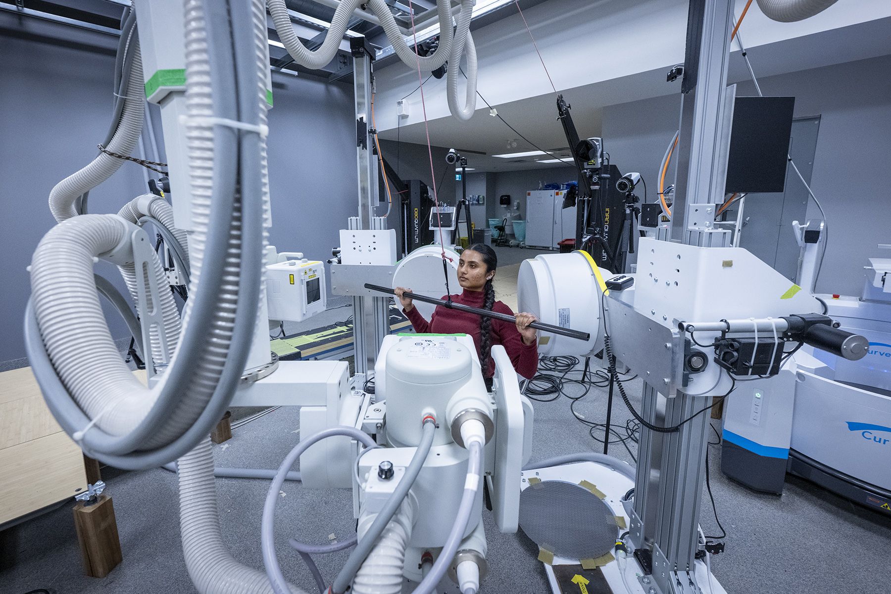
Biplanar videoradiography (BVR) allows researchers to see shoulder bones move inside the body in real-time.
Researchers at Kingston Health Sciences Centre (KHSC) and Smith Engineering at Queen’s University have used advanced imaging technology to debunk long-held assumptions about how the human shoulder moves. The study is the first of its kind in Canada and represents a major step forward in understanding shoulder health and injury, insights which could one day help improve rehabilitation therapy, injury prevention, and design more effective surgical procedures.
The study, published in the Journal of Biomechanics, was conducted in the Skeletal Observation Laboratory (SOL) at located at KHSC’s Hotel Dieu Hospital site, a joint initiative of Smith Engineering and the KHSC Research Institute (KHSC-RI).
Using biplanar videoradiography (BVR), a specialized X-ray system, researchers were able to see shoulder bones moving inside the body in real-time, with high accuracy. Unlike traditional X-rays, which show a single still image, this technology captures how joints move during activities like lifting, pushing and pulling. The X-ray images are then combined with 3D bone mapping from CT scans, creating a 3D image of the shoulder joint in motion.
“Previous research shows that specific movement patterns are associated with shoulder pain. However, most of this research has looked at people who already have pain, which makes it hard to know what’s cause and what’s effect,” says Erin Lee, first author of the study, former PHD student at Queen’s and current postdoctoral fellow at the University of Waterloo. “We wanted to understand how a healthy shoulder moves before pain or injury begins.”

X-ray images are combined with 3D bone mapping from CT scans to create a 3D image of the shoulder
The research team studied healthy volunteers as they performed two different arm movements (pushing and pulling) that placed opposing forces on the shoulder. The researchers saw that even when the arm reached the same position, the shoulder blade moved differently depending on what movement the person was trying to manage.
It had long been assumed that more upwards movement of the shoulder blade was always better. Instead, the findings suggest the shoulder blade subtly adjusts its motion to keep the joint stable during different demands.
“This kind of research allows us to revisit long-standing assumptions about how the human body works. Many of those assumptions have shaped treatment strategies, yet they were made before we could directly measure skeletal motion. Being able to do that helps us understand how the body is meant to function,” said Michael Rainbow, director of the Skeletal Observation Laboratory and associate professor of Mechanical and Materials Engineering. “This kind of study is essential for designing better treatments in the future. We’re proud to operate a truly unique research space here in Kingston as our lab is one of only two like it in the country.”

Michael Rainbow (L) with a group of students from the Skeletal Observation Lab
The Skeletal Observation Laboratory demonstrates the close partnership between Queen’s University and the hospital, bringing together researchers, surgeons, imaging experts, and engineers to use their skills to improve understanding of the human body and future patient outcomes.
“We see the impact of this kind of research when medical teams are able to work side-by-side with researchers to advance our knowledge of the human body,” says Dr. Stephen Smith, president and CEO of the KHSC-RI and deputy vice-principal research at Queen's. “By creating shared spaces where medical teams and engineers are able to work together, our patients are the ones who benefit directly from the information we uncover.”
To learn more about the Skeletal Observation Laboratory visit: https://skeletalobservationlaboratory.com/


